Restrictions: All visitors are required to wear a mask or face cover to enter. Please do not visit if you feel unwell (cough, stuffy nose, fever or sore throat)
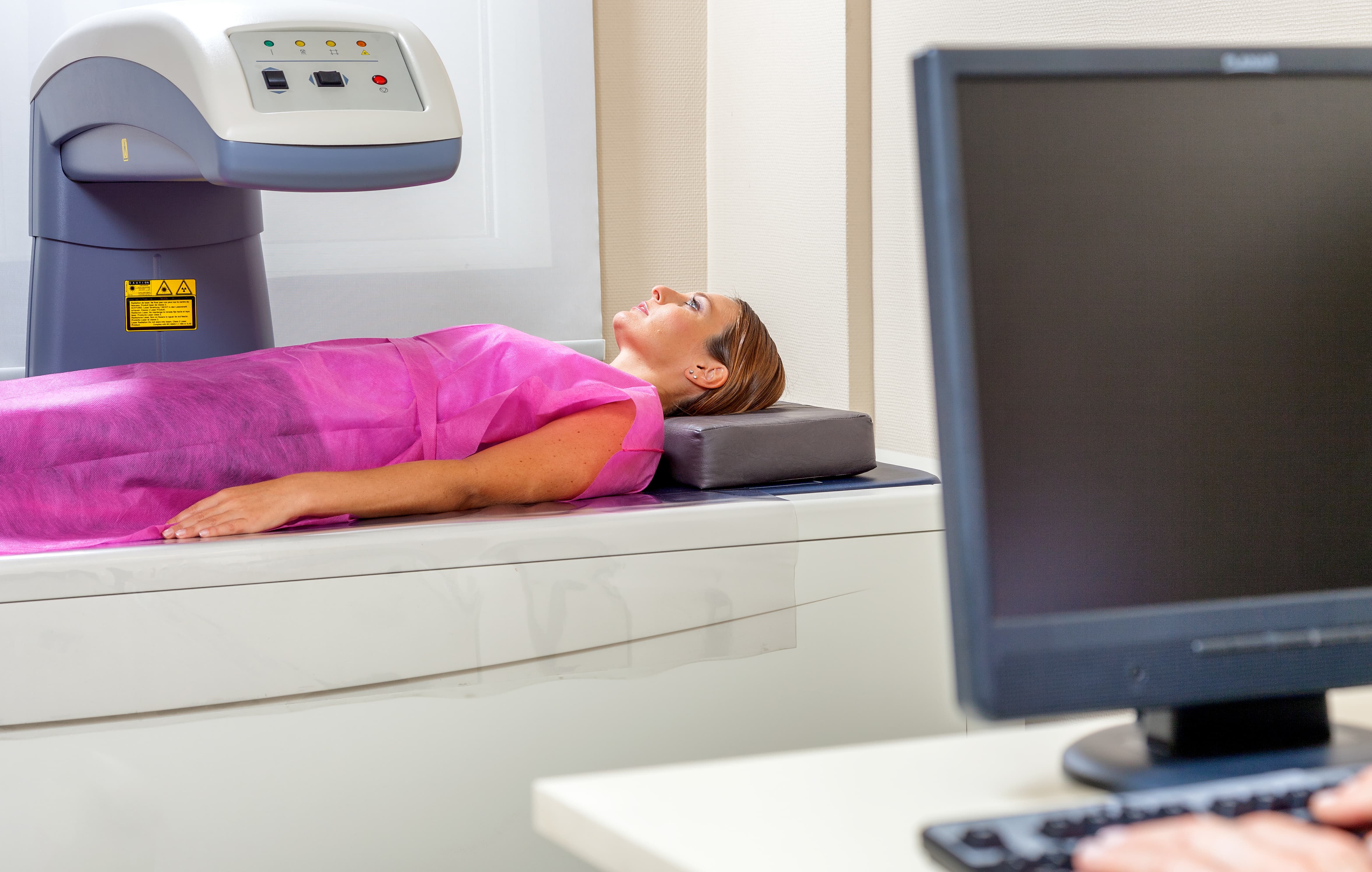
Bone Density Test DEXA & DXA
Bone Density Scan (DEXA scan, DXA scan)
A bone density scan or DEXA scan or DXA scan is both non-invasive and pain-free. This test determines the strength of your bones by measuring the grams of calcium and other bone minerals.
Request an Appointment

Symptoms and Diagnosis
Screening mammography.
This test will report the likelihood of developing osteoporosis, a disease of fragile and weak bone and occasionally even find osteoporosis related fractures that have already occurred.
This Scan will help to:
Assess and identify decreases in bone density before you break a bone
Determine your risk of broken bones (fractures)
Confirm a diagnosis of osteoporosis
Monitor osteoporosis treatment
You may be referred to have this test if you have:
Lost height
Fractured a bone
Taken certain drugs
Received a transplant
Had a drop in hormone levels
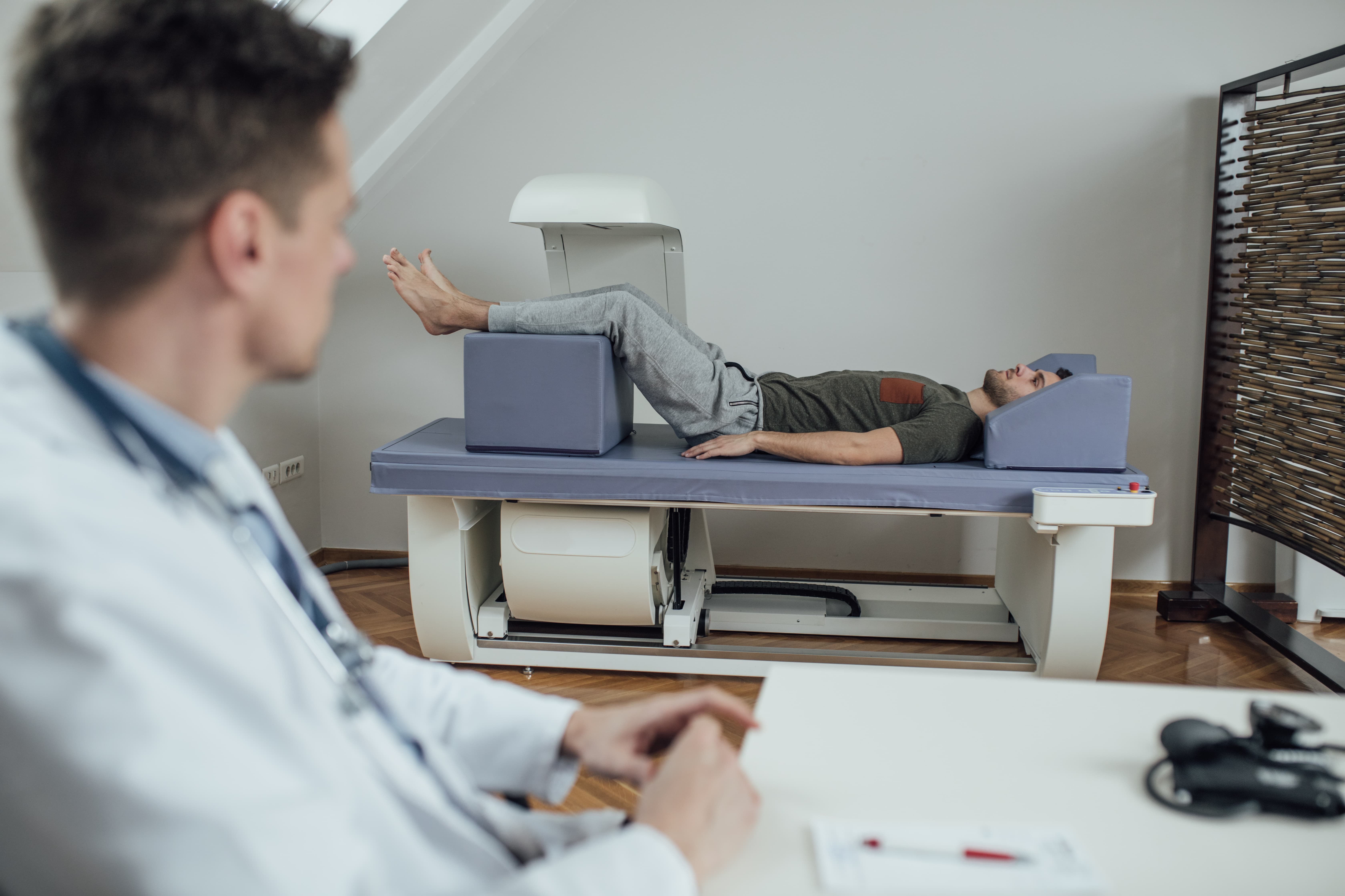
What to expect at your Bone Density Scan appointment.
Bone density scans require minimal and brief preparation. If you’ve recently had a barium exam, had contrast material injected for a CT scan, or nuclear medicine test, ensure you let the clinic know. The amount of radiation you’re exposed to is extremely low.
How often to repeat a Bone Density Test or DEXA Test, DXA Test.
If you are taking osteoporosis medicine you should repeat a bone density test by CDN every 1-2 years. You should repeat your bone density test every year after starting osteoporosis medication.
Before Your Scan
Before the bone density scan, you may be asked to wear a paper gown. Do not take calcium supplements for at least 24 hours before your bone density test. Patients should be advised not to wear clothing with buttons or zippers or snaps.
NOT to be booked within 10 days of a Barium study or within 7 days of a Nuclear Medicine/ X-Ray Dye study. Bring a list of your meditations.

During The Scan
An x-ray machine will scan those areas where bone degradation is most commonly found including the back and hips with minimal physical contact.
After The Scan
Once the test is complete you will be able to return to your normal activities.
Results
The test will be interpreted by a radiologist and the results will be communicated to your referring doctor. You may discuss the results of the exam with your doctor at a follow-up appointment.

Locations of the Bone Density Scan
-
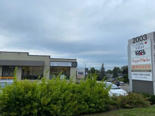
Orleans Imaging
2003 St Joseph Blvd, Orleans, Ontario
-
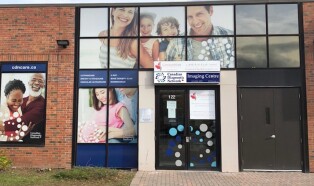
Kanata Imaging
150 Katimavik Rd , Unit 122, Ontario (Located Behind the Building)
-
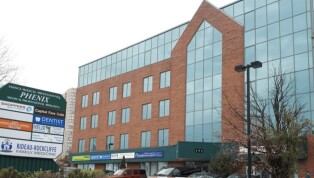
Phenix Imaging
595 Montréal Rd, Suite 205, Ottawa, Ontario
-
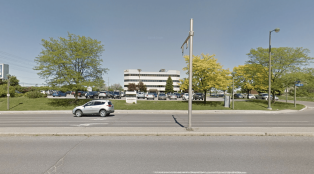
Nepean Imaging
1 Centrepointe Dr, Unit 106, Nepean, Ontario
-
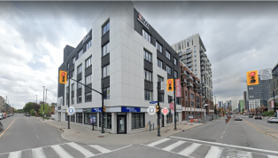
Charlotte Imaging
168 Charlotte St , Suite 302, Ottawa, Ontario
-
Barrhaven Imaging
605 Longfields Dr , Ottawa, Ontario
Make your Appointment
Your examination or doctor’s visit is 4 easy steps away
Request an Appointment
You can also be interested in
-

Mammography
Detects breast anomalies via low-dose X-rays.
-

Ultrasound
Sound waves for real-time internal imaging.
-

X-Ray
Non-invasive and pain free method of diagnostic imaging.
-

Vascular ultrasound
Sound waves map blood flow in veins and arteries.
-

ABU & 3D Ultrasound
Detailed organ imaging with 3D abdominal views.
-
Breast Ultrasound
Uses sound waves to examine and detect potential breast problems.
-

Echocardiography
Also known as a cardiac ultrasound, is a non-invasive imaging technique used to assess the structure and function of the heart.
-

Body Composition Analysis
Precision body analysis for informed health choices.